Low-Dose 3D imaging:
less radiation, more information
Downtown Dental believes in responsible radiation practices for patients. Dental x-rays are often the only way to diagnose disease and conditions that are not evident in a visual exam. Identification of cysts, cancerous and non-cancerous tumors, tooth decay (cavities), and Periodontal disease is often only possible with diagnostic x-rays. In addition to saving you money on treatment by early diagnosis, x-rays identifying cancerous lesions can save your life.
At Downtown Dental, we only utilize the latest low dose digital 2D and 3D CBCT technology. With the latest in 3D imaging, our clinicians can see more, with even less radiation than ever before. This allows us to provide you with the essential information you need to make the best decisions about your healthcare.
Best of all, 3D imaging emits an extremely limited and safe amount of radiation. A LOW DOSE 3D CBCT scan exposes the patient to LESS radiation than a traditional ‘full mouth’ series of traditional 2D x-rays routinely done in general dental practices.

We use our 3D imaging for cosmetic/restorative cases, root canals, and dental implants. We do not blindly take routine x-rays, either: we consider both 2D and 3D imaging against your risk factors, and we consider what is the lowest risk radiation in order to gain the best and most actionable information. Downtown Dental adopts the same approach used in modern medicine, we ensure we are prescribing imaging to achieve patient health with radiation doses as low as reasonably achievable (ALARA).
What is the benefit for our patients?
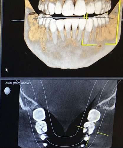
Diagnostic Accuracy
2D imaging captures information and then compresses that information into 2 dimensions. So problems must be relatively far progressed before there is enough depth of changes in the bone and tooth structure to be seen in a 2D radiograph. A 3D image does not have those limitations, so problems can be diagnosed when they are much less advanced. The sooner the diagnosis, the sooner you can resolve the issue. Many infections in bone or cavitations cannot be easily visualized with traditional 2D imaging but are easily visible in 3D imaging.
Minimal Radiation Exposure
Our digital X-ray technology lessens the radiation exposure of previous x-rays by up to 90%!
Non-Intrusive
Most of our imaging can be done by standing in our Planmeca Promax machine and letting the equipment rotate around your head. No more mouth full of sensors hurting the roof or floor of your mouth.
Visualize Anatomy
We have the ability to visualize many parts of anatomy not visible in traditional dental imaging. Our imaging can be used by your ENT or primary care physician to assess potential causes for breathing issues and chronic sinus infection. Our imaging can also be used to visualize the upper cervical neck anatomy in order to assist in diagnosis of conditions causing neck and craniofacial pain. Our imaging also allows us to visualize anatomical markers to monitor growth and remodeling in children and adults.
More Effective Treatment Plans
Less uncertainty means creating better, more effective treatment plans and more accuracy in assessing the benefits and risks of treatment options.